Anatomy Muscles Pelvis ~ Monmed Pelvic Model 6pc Life Size Anatomical Female Pelvis Model With Muscles Walmart Com Walmart Com. Psoas consists of a pair of deep muscles (psoas major and iliacus) located on each side of the pelvis in the abdomen. It is usually divided into two separate anatomic regions: These bones connect the axial skeleton to the lower limbs, and therefore play a role in bearing the weight of the upper body. The hip bone has three parts: There are three bones of the pelvis:
Hip and thigh anatomy | pelvis, pelvic bone, pelvis anatomy / learn about anatomy muscles pelvis with free interactive flashcards. The pelvis consists of the sacrum, the coccyx, the ischium, the ilium, and the pubis. Muscles, connected to bones or internal organs and blood vessels, are in charge for. Pelvic floor anatomy springerlink : The pubic symphysis and the sacroiliac joint, and reinforced by pelvic muscles.
Female Pelvic Floor 1 Anatomy And Pathophysiology Nursing Times from cdn.ps.emap.com They are also known as the inner hip muscles and deep external rotators. The muscles of the pelvis, hip and buttock anatomical chart shows how each muscle in this area of the body works with the others, and the various minor systems within the major ones. The ilium, ischium and the pubic bone. The muscles of the pelvic floor are collectively referred to as the levator ani and coccygeus muscles. The pelvic floor muscles provide foundational support for the intestines and bladder. The pelvic region holds major organs under its layers of muscles. The pelvis is the lower portion of the trunk, located between the abdomen and the lower limbs. The muscles of the pelvis and hip control the vast range of movement of the legs and torso.
The hip bone has three parts:
Below the sacrum is the coccyx, or tailbone, a section of fused bone that is the end of the vertebral. The pelvis is the lower portion of the trunk, located between the abdomen and the lower limbs. Ventral primary rami s4 and s5. These muscles, including the gluteus maximus and the hamstrings, extend the thigh at the hip in support of the body's weight and propulsion. Use the mouse scroll wheel to move the images up and down alternatively use the tiny arrows (>>) on both side of the image to move the images.>>) on both side of the image to move the images. It helps maintain erect posture, abducts the thigh, and rotates the thigh outward. This mri male pelvis axial cross sectional anatomy tool is absolutely free to use. The pubic symphysis and the sacroiliac joint, and reinforced by pelvic muscles. The muscles of the pelvis and hip control the vast range of movement of the legs and torso. These bones connect the axial skeleton to the lower limbs, and therefore play a role in bearing the weight of the upper body. There are four articulations within the pelvis: The floor of the pelvis is made up of the muscles of the pelvis, which support its. Related posts of muscle anatomy pelvis muscle anatomy coloring pages.
Acts with levator ani muscle to support pelvic contents and is important for micturition and defecation. Some conditions that can affect the female pelvis. The pubic symphysis and the sacroiliac joint, and reinforced by pelvic muscles. Large ligaments, tendons, and muscles around the hip joint hold the bones (ball and socket) in place and keep it from dislocating. Hip and thigh anatomy | pelvis, pelvic bone, pelvis anatomy / learn about anatomy muscles pelvis with free interactive flashcards.
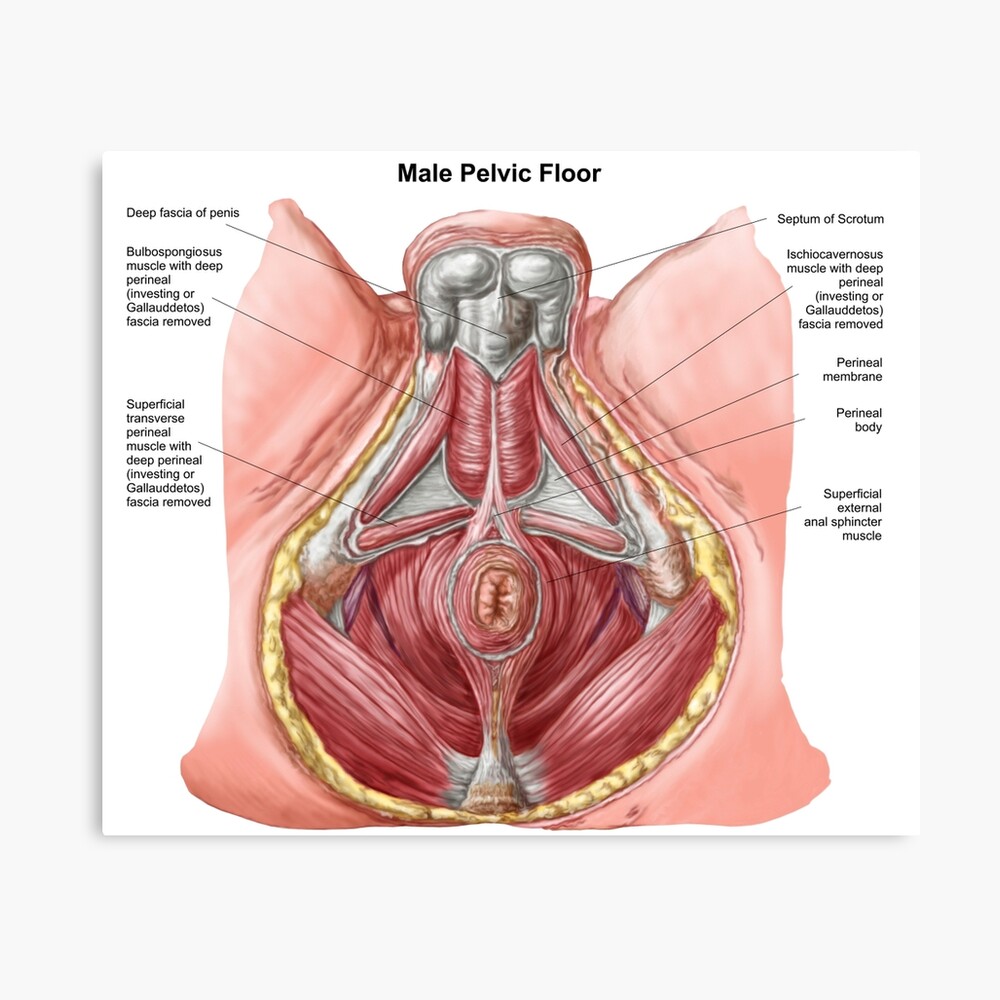
Pelvic Floor Of Human Male Poster By Stocktrekimages Redbubble from ih1.redbubble.net Ventral primary rami s4 and s5. Use the mouse scroll wheel to move the images up and down alternatively use the tiny arrows (>>) on both side of the image to move the images.>>) on both side of the image to move the images. Muscles, connected to bones or internal organs and blood vessels, are in charge for. The pc muscle acts as the queen bee of the pelvic floor so when the pc muscle contracts, all the other muscles and sphincters follow along. There are three bones of the pelvis: On the posterior side they are the glutei and on the anterior side the hip muscles extending into the thighs. There are four articulations within the pelvis: It can be described as one of the bodies diaphragms.
Acts with levator ani muscle to support pelvic contents and is important for micturition and defecation.
Attached to the pelvis are muscles of the buttocks, the lower back, and the thighs. These muscles have attachments to the pelvis as follows. The pc muscle acts as the queen bee of the pelvic floor so when the pc muscle contracts, all the other muscles and sphincters follow along. The muscles of the pelvic floor are collectively referred to as the levator ani and coccygeus muscles. The pelvic cavity opens superiorly to, and is continuous with, the abdominal cavity through the pelvic inlet. Psoas consists of a pair of deep muscles (psoas major and iliacus) located on each side of the pelvis in the abdomen. Large ligaments, tendons, and muscles around the hip joint hold the bones (ball and socket) in place and keep it from dislocating. The pelvic floor muscles include; The hip joint is one of the most flexible joints in the entire human body. Related posts of muscle anatomy pelvis muscle anatomy coloring pages. Hip and thigh anatomy | pelvis, pelvic bone, pelvis anatomy / learn about anatomy muscles pelvis with free interactive flashcards. These bones also act as attachments for many muscles and ligaments within the pelvis and lower limbs. Use the mouse scroll wheel to move the images up and down alternatively use the tiny arrows (>>) on both side of the image to move the images.>>) on both side of the image to move the images.
The bony framework of the pelvis is called the pelvic girdle.it is composed of the two hip bones and the sacrum. The pelvis contains a large number of organs, bones, muscles, and ligaments, so many conditions can affect the entire pelvis or parts within it. The small intestine is the longest part of the. The pelvic floor or pelvic diaphragm is composed of muscle fibers of the levator ani, the coccygeus muscle, and associated connective tissue which span the area underneath the pelvis. The pc muscle acts as the queen bee of the pelvic floor so when the pc muscle contracts, all the other muscles and sphincters follow along.
3d Rendered Illustration Of Pelvic Muscles Anatomy Canstock from comps.canstockphoto.com The pelvis consists of the sacrum, the coccyx, the ischium, the ilium, and the pubis. Below the gluteus maximus is the smaller gluteus medius. It helps maintain erect posture, abducts the thigh, and rotates the thigh outward. The pelvis is the lower portion of the trunk, located between the abdomen and the lower limbs. Some conditions that can affect the female pelvis. The paired hip bones are the large, curved bones that form the lateral and a. The four groups are the anterior group, the posterior group, adductor group. The pubic symphysis and the sacroiliac joint, and reinforced by pelvic muscles.
It is usually divided into two separate anatomic regions:
The small intestine is the longest part of the. The pelvis contains a large number of organs, bones, muscles, and ligaments, so many conditions can affect the entire pelvis or parts within it. The pelvis consists of the sacrum, the coccyx, the ischium, the ilium, and the pubis. The bony pelvis consists of the two hip bones (also known as innominate or pelvic bones), the sacrum and the coccyx. The largest of them is the most superficial muscle, the gluteus maximus. Below the gluteus maximus is the smaller gluteus medius. The sacrum, five fused vertebral bones, joins the pelvis between the crests of the ilium. Small and deep muscles which mainly externally rotate the thigh at the hip joint and stabilize the pelvis. These bones connect the axial skeleton to the lower limbs, and therefore play a role in bearing the weight of the upper body. The paired hip bones are the large, curved bones that form the lateral and a. There are three bones of the pelvis: Attached to the pelvis are muscles of the buttocks, the lower back, and the thighs. Muscles that attach from the pelvis to the trunk and cross the lumbosacral joint muscles that attach from the pelvis to the thigh/leg and cross the hip joint pelvic floor muscles that are located wholly within the pelvis